
Coming into Focus Radiology & Imaging Introduces Digital Mammography
Perhaps no high-tech item has captivated America over the past few years like the digital camera. The devices have become so popular, in fact, that shooting on film seems almost quaint.
In the same way, many doctors expect that digital imaging will eventually supplant film for medical purposes, although it’s taking a more circuitous route to widespread implementation, due in part to cost. But it’s a technology in which Radiology & Imaging (R&I), a network of medical imaging services in the Pioneer Valley, finds itself ahead of the curve.
The company recently introduced digital mammography to its roster of women’s services — the first facility in the region to make the transition. Digital mammography replaces X-ray film with detectors that convert X-rays into electric signals, similar to the way digital cameras work. These signals are used to produce images of the breast that can be seen on a computer screen or printed on special film to look like regular mammograms.
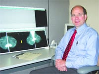
“What it does is, instead of using film to obtain a mammography image, the digital capture allows images to be taken more precisely,” said Dr. David March, R&I’s director of breast imaging. “After they’re taken, the contrast can be changed, and images can be enlarged or inverted. You can do a lot with digital images that you can’t do with film.”
The patient doesn’t notice anything different during the exam, however. From her point of view, digital mammography is conducted essentially the same way as the screen-film version. But to the medical professional who is reading the image, the difference can be stark.
“With film mammography, you’re basically limited by the quality of the film that comes out of the processor. You can’t do image manipulation,” March said. For example, increasing the contrast of a digital image can make it easier to detect malignancies earlier — and early detection is, of course, the universal goal of cancer prevention.
Following the Science
March said the company made the decision to go digital following a report published in the New England Journal of Medicine. In that study, more than 49,000 women were tested with both film and digital mammography, and the results were independently reviewed.
“There were three groups of women for whom digital mammography was significantly better at identifying cancer,” March said; these groups included women under age 50, pre-menopausal and peri-menopausal women, and those with dense breast tissue. “This was the first study that showed a definite advantage for digital mammography in certain groups of women, and that’s what made us decide to switch to digital.”
Constantine Gatsonis, professor of Community Health and Applied Mathematics at Brown University and one of the authors of the report, said the study provides some of the best data ever gathered on the diagnostic accuracy of mammograms.
“The data show that digital mammography is, on average, as good at detecting breast cancer as film mammography — and, in some important subgroups of women, digital performs even better,” Gatsonis said. She added, however, that “neither film nor digital mammography is able to catch every cancer. So this study data can be used to develop and improve mammography in the coming years.”
Digital mammography functions with computer-aided detection, or CAD, a diagnostic tool used by the radiologist during the interpretation of the mammogram. March said the use of CAD has been reported to increase breast cancer detection by up to 20{06cf2b9696b159f874511d23dbc893eb1ac83014175ed30550cfff22781411e5}.
Radiology & Imaging is also setting itself apart by using what is known as “full-field” digital mammography. The specific equipment, called the Selenia system and produced by Bedford-based Hologic, boasts the largest available field of view, and can image most patients, including larger women, without the need for multiple images or repositioning of the equipment.
“Some digital units produce images that are smaller than the size you need to accommodate many women, so a lot of overlapping images have to be obtained to cover all the tissue,” March said.
“It makes the images more difficult to interpret because you’re doing redundant readings on a lot of overlapping areas,” he explained. “The system we chose has the largest field of view available, so it really cuts down on the need for extra views.”
Getting the Picture
Radiology & Imaging has installed the new technology at both its Springfield locations, on Liberty Street and Medical Center Drive, as well as its Enfield, Conn. location. All three sites also offer an array of other imaging services in the company’s breast imaging and diagnostic program.
“For breast imaging, we offer state-of-the-art equipment for a high-resolution ultrasound, and we also have stereotactic biopsy equipment that’s all digital,” March said. “We do a large number of biopsies on findings from mammograms or ultrasound; we can use either modality to guide biopsies.”
In fact, March said, Radiology & Imaging was the first facility in Western Mass. to perform core needle biopsy using these image-guidance systems —a combination of technologies that give women a more comfortable experience, which in turn reduces the fear of getting potential problems checked out.
“The good thing is that most biopsies turn out to be benign,” March said. “And these biopsies with image-guidance systems are shown to be extremely accurate and well-tolerated by women.”
Radiology & Imaging also offers ultrasound services for other parts of the body, as well as bone densitometry. Meanwhile, it remains the only center in the region to offer digital mammography, although Baystate Health is planning to add it to its roster of imaging services soon.
“Suddenly, the science was there,” March said of the decision to move to digital mammography. “We decided it was time, that this was something we should do now.”
It’s a decision whose rationale is coming into focus — and benefiting women across the Pioneer Valley — one image at a time.

Comments are closed.