
Five Heartbeats In That Short Time, Mercy’s 64-slice CT Scanner Will Capture Your Heart
In the early days of photography, a person had to sit still in front of a camera for a long period of time, unmoving, to produce a clear, non-blurry portrait. In fact, one of the earliest photographs, an image of a building taken in 1826, took eight hours to expose, and has sunlight visible on both sides of the building.
Clearly, action shots were out of the question.
Today, high-speed, auto-focus digital cameras allow anyone with no photography experience at all to capture perfectly sharp images, even of objects in motion, in a split second with the push of a button.
Radiologists have experienced a similar quantum leap in technology, as increasingly high-speed CT (computed tomography) scans are producing three-dimensional images of the inside of the body with a crispness and clarity that were unthinkable just a decade or two ago.
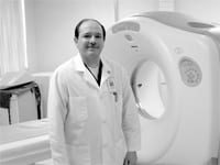
Mercy Medical Center in Springfield is one of the hospitals using the latest CT technology, what’s known as a 64-slice scanner, said Carlos Valdez, the hospital’s chief of Radiology and Cardiac Radiology. “Because it’s faster, the patients like it,” he said, “and for us, we’ve improved the quality, and the pictures are crisper.”
Rapid Advances
Computed tomography is a medical imaging method which uses a series of two-dimensional X-ray images to generate a three-dimensional image of the body’s internal organs. But whereas the earliest CT scans were often too slow to produce clear, accurate images — especially of an organ in motion, like the heart — they were limited in their effectiveness.
“There has been a progression in the technology over the past 10 years or so,” Valdez explained, noting that the earliest CT scanners had just one ‘detector,’ the imaging device that takes the X-ray pictures as it rotates around the body; that image is called a ‘slice,’ and a series of slices can be assembled to produce a 3D picture of the body on a computer screen.
During the past decade, the technology advanced to four-slice scans, which took four slices per rotation, speeding up the process and producing more accurate images. But the heart remained a problem, because it moves when it beats, and radiologists taking even four slices at a time found it difficult to produce high-quality images of the heart. “The body is in constant motion, and one of the things that’s always moving is the heart,” Valdez said.
“Initially, you went around the patient once and had one slice; then you got four slices every time you went around,” he explained, adding that 16-slice scanners followed about five years ago. “The latest generation, the 64-slice machine, is of course much faster. By taking the images 64 times faster, if allows you to freeze the motion inside of the body.”
The high-speed scan also allows radiologists to examine the fine blood vessels around the coronary arteries, a process which previously required a catheter strung through the groin into the chest, because the previous CT scan speeds were not sufficient to create an accurate image from the outside, Valdez said.
“We can take pictures of the entire heart in just five heartbeats,” he noted. “In just a few seconds, we have pictures of all the vessels in the heart, and with much better-quality images compared to what we used to get. That’s just amazing, and it’s becoming very useful in looking for diseases of the heart.”
He stressed, however, that the 64-slice CT is not used to check for blockages in a potential cardiac arrest situation. “If a patient is in pain or sweating, he needs an arteriogram, by which we can also put a stent in the vessel and treat the blockage,” Valdez said. “If you do a CT scan and find that the patient still needs a stent, you’ve wasted time.
“But for someone who’s not having immediate pain,” he explained, “perhaps someone with a family history for coronary artery disease, it’s a good test to tell whether the symptoms a patient has had are related to heart disease.”
Body of Evidence
Valdez was quick to note that the state-of-the-art scanner has improved CT images well beyond the heart.
“We can scan every part of the body faster than before, from the brain to the chest, the pelvis, and the bones of the hands and the feet,” Valdez said, noting that the improvements in brain scans aren’t so dramatic as those of the heart, but “there’s a set of small vessels in the brain that feed the brain, and they can be scanned very nicely with the 64-slice.”
Other benefits have emerged as well. For example, a contrast die is normally injected into an artery to help radiologists determine the level of blockage in the passageway. “With a quicker scan, we can lower the volume of the contrast to the patient, and the more contrast we use, the more likely it is to have side effects, including kidney problems,” Valdez said. “By allowing us to decrease the volume of the dye we use to enhance the vessels, it also potentially saves some money for the hospital.”
The imaging of arteries is now so accurate, Valdez explained, that a negative reading basically negates the need for an arteriogram, which saves the patient from having to undergo an additional procedure. “We can scan other parts of the body as well, like inside the abdomen and the arteries to the lower extremities,” he said. “Because those vessels are bigger than the ones in the heart and not really moving, the quality wasn’t as bad before, so the improvement is not as great — but it’s still an improvement.”
And those advances haven’t ended yet.
“They’re working on a 256-slice CT,” Valdez said. “The technology never stops. Computers become obsolete three or four years after you buy them, and the same thing is happening with CT scanners. They’re not obsolete, exactly, but the improvement is so great that you just want to get the next generation instead of keeping the earlier one.”
