The Latest MRI Software is Saving Lives at Holyoke Medical Center
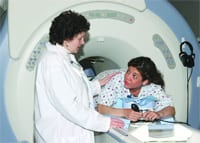
HOLYOKE — Holyoke Medical Cen-ter’s top-of-the-line Magnetic Resonance Imaging (MRI) machine is helping to save patients’ lives every day, while exceeding physicians’ expectations by producing the clearest, highest-resolution images time and time again.
Fueled with the latest software technology, the General Electric ‘1.5 Signa EXCITE High Definition MRI Scanner’ captures images of the brain, cervical spine, neck, lumbar spine, knees, shoulders, and of the vascular system (blood vessels) better than ever before.
With its multi-planar imaging capability, high-resolution magnet, and high definition, multi-channel coils, radiologists are able to detect very small brain tumors, perform breast biopsies, diagnose liver and abdominal cancers, and locate arterial stenosis much earlier than ever before.
This technology lends itself to earlier detection, faster treatment, and resolution of the disease, resulting in better prognosis for patients throughout the Greater Holyoke community and beyond, said HMC Chief of Radiology Dr. Mrinal Mali.
“This MRI is still the best modality around. It’s the latest machine available in the 1.5 Tesla platform. We are using new sequences to scan the whole body from the abdomen to the feet and to diagnose problems in the upper and lower extremities,” said Mali. “For arterial narrowing, including toes and palms, looking for the smallest vessel has tremendous gain. Evaluation of the ear and eye has never been better. We’re picking up smaller tumors. Even the resolution in the brain for looking at smaller arteries is much better.”
One of the biggest advantages to the EXCITE MRI in vascular imaging is that images are taken every second, in real time with a software option called ‘TRICKS.’ Whether imaging blood vessels of the feet, the heart, neck, carotid arteries, renal arteries, kidneys, or the brain, the MRI allows for faster, more-accurate diagnosis.
“Previously, a patient having a conventional angiograpy would have a catheter threaded into the thigh to inject contrast into the blood vessel. An MRA (MR Angiography) is a non-invasive test that yields the same results,” said MRI Supervisor Nancy A. King. “This procedure only requires an intravenous injection with a small needle in the arm. With our new MRI technology, we’re able to achieve complete separation of arterial and venous phases with one injection. This is a huge advance.”
In brain imaging, new software called ‘PROPELLOR’ allows physicians to capture very clear images, even if a patient moves his head during the scan. This is an asset for fidgety children as well as patients who tremor from Parkinson’s Disease. In breast imaging, the MRI is capable of scanning three-dimensional images of both breasts in a single patient visit using software called ‘VIBRANT,’ and can even be used to perform breast biopsies. The MRI excels at imaging the abdominal region, using a sequence called ‘LAVA’ to scan an entire region of liver, in one breath hold, with high in-plain resolution and high asset factors, said Mali.
While it used to require 60 seconds of breath-holding to capture accurate, multi-dimensional images of the liver, the EXCITE MRI can capture 100 images in one 20-second breath hold, he said.
Mali depends upon the MRI — an average scan lasts about 30 minutes depending on the exam — to help him diagnose a variety of diseases.
“For me, I use MR for everything and there are endless possibilities including a patient with headaches, stroke, breast lesions or with family history who is at high risk for breast cancer,” said Mali. “It can also be used for patients with peripheral vascular disease (leg pain, clotting), any abdominal masses, any nervous system issues. This is the best modality we have.”
HMC’s MRI Unit is open Monday through Friday from 7 a.m. to 11 p.m. and Saturday from 7 a.m. to 4 p.m. and is located conveniently on the first floor, just down the hall from the Laboratory and adjacent to the Blood Bank.


Comments are closed.