Vascular Ultrasound: Easy And Risk-free Ultrasound Can Detect Developing Blockages And Narrowing In The Veins And Arteries
Ultrasound machines are probably most famous for giving parents-to-be a joyful first glimpse of their unborn baby.
But Holyoke Medical Center is also at the forefront of using vascular ultrasound to study blood flow through the veins and arteries to determine whether there are potentially dangerous blockages or narrowing.
Michael A. Zwirko, vice president of Operations, said HMC opened its Vascular Ultrasound Laboratory last year to respond to an urgent and growing need within the community for the procedure.
“Given our patient population and the prevalence of arterial sclerotic disease in our community and in patients living longer, we are seeing more of a need to do more vascular procedures,” Zwirko said. “We needed to have a vascular lab. We started buying equipment and training our technicians. We set up a separate area for the lab and started concentrating on providing high quality vascular ultrasound techniques.”
Prior to the establishment of the Vascular Ultrasound Laboratory, patients from the Holyoke area had to travel to Springfield, Hartford, or Worcester to have procedures done. Now, HMC’s lab makes access to these sophisticated tests more convenient. Once a diagnosis is made, follow-up treatment is also available at HMC.
“It’s inexpensive and non-invasive,” said Dr. Jack Horky, HMC director of Vascular Interventional Radiology. “It’s easy, comfortable and risk-free for the patient. If we identify a disease using vascular ultrasound, we can then go onto do MRI (magnetic resonance imaging), a CT (computed tomography) scan, or an angiogram here at HMC.”
Blockage or narrowing of veins and arteries can lead to serious health problems. First, it can be quite painful to walk for a person suffering from circulation problems in the legs. In the more severe cases, the condition can cause tissue breakdown and in some instances lead to amputation.
Narrowing or blockage of veins and arteries leading to the brain can be a precursor to stroke.
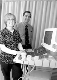
Vascular ultrasound is as safe and painless as any other ultrasound procedure. The patient lies down on an examination table and the sonographer applies a gel to the areas of the legs or neck to be examined. The sonographer then sweeps an ultrasound probe, called a translucer, over those areas. Images are projected onto the ultrasound monitor and the sonographer occasionally stops to take a picture of an image.
HMC has three key pieces of equipment in the Vascular Ultrasound Laboratory. The HDI 5000 ultrasound uses Doppler technology to measure the speed of blood flowing through the veins.
“Blood flow will increase in speed in an area of narrowing,” Horky said.
Vascular Surgeon Dr. Robert A. Goodman last year donated an ImexLab 9000, a plethysmograph that measures blood pressure in the legs. This procedure is similar to a routine blood pressure exam, where cuffs are wrapped around the patient’s legs and arms. The cuffs are then inflated and pulse waves are measured from each cuff. A decrease in pulse waves may indicate an area of blockage.
The third piece of equipment used in the lab is a treadmill. Horky said exercising on a treadmill may help reveal hidden areas of blockage. A patient may exercise on the treadmill before a plethysmograph examination. +
“Sometimes tests will come back normal, but the patient still says the legs hurt after walking,” Horky said. “Exercise will help uncover the problem.”
Sonographer Melanie S. Krol, RDMS, RVT, said vascular physicians will refer their patients to the Vascular Ultrasound Laboratory after seeing conditions that could mean circulation problems, such as pain after walking, leg cramps severe enough to keep a patient awake at night, or chronic cold feet and toes.
“Using the vascular ultrasound we can diagnose the condition and that allows the physician to know if the patient needs surgery or other kind of treatment,” said Krol, who is supervisor of Vascular Ultrasound. “A patient’s condition shows up right away on the screen. A serious condition goes right to the physician’s office many times before the patient leaves the hospital.”
Goodman pointed out that physicians rely heavily on vascular ultrasound.
“The tests are 96{06cf2b9696b159f874511d23dbc893eb1ac83014175ed30550cfff22781411e5} accurate,” he said. “We don’t have to do invasive procedures because the tests are so accurate. Many vascular surgeons, including myself, are now operating based on the results of the ultrasound. The ultrasound is so accurate we can plan surgery based on its results.”


Comments are closed.