
Holyoke Medical Center Introduces Its State-of-the-art ‘16-slice’ CT Scanner
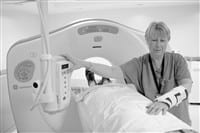
HOLYOKE — Standing center-stage inside a spacious, freshly-painted new suite at Holyoke Medical Center (HMC)’s Radiology Department is one of the newest, most sophisticated 16-slice CT scanners available.
The $1.3 million scanner, manufactured by General Electric, not only collects very high resolution, cross-sectional images in a fraction of the time of less capable machines, but its state-of-the-art computer software allows radiologists to reconstruct internal organs and reconfigure their positions for a better view of blood flow and disease processes.
To understand how the 16-slice CT scanner works, picture a single Slinky coiled around a patient’s upper torso. Now, picture 16 Slinkies wrapped around the same section of the anatomy, placed much closer together and with significantly less space between each ring, allowing for incredibly clear, and extremely detailed, three-dimensional images.
This is the difference between Holyoke Medical Center’s single-slice CT scanner, which will still be used for routine CT scans in the Radiology department, and the new, 16-slice CT scanner that makes its home inside the new, state-of-the-art CT suite.
“We just jumped right in with both feet and are amazed at this machine’s capabilities and how much more efficient things will be here in Radiology, never mind how much faster and more accurate the results will be for patients requiring CT scans,” said HMC Special Imaging Supervisor Laura Maziarz.
The 16-slice CT scanner can scan a patient’s body from the chest to the feet in just 40 seconds, a procedure that previously could have taken several minutes. It can scan the chest in nine seconds, with one breath hold. Because it is so fast and accurate, patients can be scheduled for CT scans every 15 minutes rather than the traditional 45-minute spacing between appointments, said Maziarz.
In addition, the scanner features a dual-head “contrast injector,” explained HMC Chief of Radiology Dr. Eyal Morag, which enhances its diagnostic capabilities as it features “perfect time enhancement phases” of blood vessels and internal organs.
This means that the contrast fluid injected literally “lights up” the body’s blood flow, clearly illustrating places with improper flow at that exact moment, he explained.
“It also enables us to administer less contrast to the patient, which is a safer thing to do,” he said.
“The exquisite capabilities of this CT scanner bring Holyoke Medical Center to an entirely new level, as far as the services that we provide for patients. Having this affects every single subspecialty in the hospital. It makes diagnosis easier and is significantly faster than other scanners, obviously resulting in more efficient, comfortable and simply, overall, a much higher quality of care for all patients,” he said.
“You can map the blood vessels supplying the brain, or the kidney, heart, lungs. The vascular work is excellent — veins, arteries, you can see if there are any aneurysms,” he continued. “You can create virtual images, you can travel inside tubular structures; it’s an incredible tool for imaging bone structures, and to evaluate hardware (surgically implanted plates or screws for example). It performs extremely accurate peripheral angiograms. That’s something you can’t do with other scanners. This is truly a tremendous machine.”
In addition, Morag said, the 16-slice CT scanner is capable of performing virtual colonoscopies (of the colon) and broncoscopies (of airways).
It can also assist dentists in reconstructions and measurements of the jaw for implants or to diagnose problems, said Maziarz, who hopes to collaborate providing this service with dentists in the Greater Holyoke region.


Comments are closed.