Breaking Point Computer Algorithm Helps Doctors Better Diagnose Fracture Risk
It’s called FRAX.
That’s short for ’fracture-risk-assessment index’, a computer algorithm that analyzes a patient’s important historical physical events (injury and sickness) and lifestyle variables (nutrition, smoking, and drinking) that can affect the quality of bones. This information is then pooled with a special scan of the skeleton for an overall analysis of bone strength during a bone-density test called bone densitometry.
And according to Dr. Robert Wool of Women’s Health Associates, an ob/gyn practice with offices in Springfield and Westfield, FRAX has changed the way he can properly diagnose his patients.
“FRAX gives me control,” he told HCN. “Instead of looking at one little piece of data, it has given me the ability to treat women with bone disease with far more accuracy, and I can make a difference; it’s revolutionized bone-health treatment.”
Because Wool’s patients are women, and natural loss of estrogen as women age through menopause is an indirect cause of bone-mass loss, they will — or already do — experience lower bone density and bone-quality issues that can lead to fractures. And these injuries are much more than an inconvenience for someone who’s fallen or the family members who have to care for them; fractures account for billions of dollars in healthcare costs, and are associated with an alarming mortality rate (more on that later).
But until the availably of the FRAX software, Wool said that no doctor or clinician had any other option than to treat an 80-year-old as he or she would a 50-year-old. That would mean medication for those who might not really need treatment, or potentially missing those who really need pharmacological therapy.
Dr. Jehangir Patel, a radiologist at Baystate Medical Center, has been instrumental in bringing an advanced bone-densitometry program to the hospital since the mid-1990s. The fight against osteoporosis and fractures has been Patel’s primary mission, especially now that the huge Baby Boomer generation is starting to hit its fragile years and experience the effects of bone disease.
Osteoporosis, Patel explained, is a bone disorder that is a result of diminished bone strength that predisposes a patient to increased risk of fracture. Bone strength has two components: bone density and bone quality.
“When we do scans, we are doing DEXA [dual energy X-ray absorptiometry], and we are really only looking at bone density,” said Patel. “In the past, there was really no good way to measure bone quality, so FRAX was developed to come up with a potential 10-year probability of fracture risk report for future hip fracture or a major osteoporatic fracture.”
According to the National Osteoporosis Foundation (NOF), FRAX was developed by the World Health Organization in collaboration with the Centre for Metabolic Bone Diseases at Sheffield University in England. FRAX integrates clinical risk factors and bone-mineral density at the femoral neck (the part of the hip bone that connects the ball joint head to the long femur bone) to calculate the 10-year probability of hip fracture and the 10-year probability of a major osteoporotic fracture (clinical spine, forearm, hip, or shoulder fracture).
As the Baby Boom generation continues to age, diseases such as low bone mass that lead to painful and mobility-limiting fractures will become more of an issue for society and the healthcare system.
For this issue and its focus on healthcare, HCN takes an in-depth look at the FRAX software and its vast potential to help prevent fratures and improve overall quality of life for individuals.
Bone Deep
According to the NOF, osteoporosis ranks with hypertension (high blood pressure), hypercholesterolemia (high LDL cholesterol), and diabetes as one of the most common chronic diseases in the U.S.
More than 10 million Americans have osteoporosis, and 33.6 million have low bone mass at the hip, the point of bone-density analysis. Using the hip as the site of base analysis is due to the extraordinary costs associated with hip fractures, said Patel, noting that 2 million fractures resulting from osteoporosis accounted for an estimated $20 billion in healthcare costs in 2010 alone. The annual cost is predicted to rise to $25.3 billion by 2025, according to the NOF.
And then, there’s the cost of human life.
“There’s a quote I read that says that a woman in her 70s or 80s who breaks her hip … there’s 25{06cf2b9696b159f874511d23dbc893eb1ac83014175ed30550cfff22781411e5} to 30{06cf2b9696b159f874511d23dbc893eb1ac83014175ed30550cfff22781411e5} mortality within that year,” said Wool. “Not because of the fracture itself, but because of other complications like bladder infections, pneumonia, lack of mobility … they get sicker because they are homebound and their quality of life goes way down and they become dependent on others.”
Wool added, “the one thing that keeps old people alive is their independence, and when they lose that, they give up their will.”
Patel said approximately 65,000 women die each year from complications from hip fracture. Of those who survive, about half are permanently incapacitated, and 20{06cf2b9696b159f874511d23dbc893eb1ac83014175ed30550cfff22781411e5} require long-term nursing home care.
An epidemiologist at the Mayo Clinic in Rochester, Minn. equated the current rates of fracture and the aging Baby Boom generation to “looking a train wreck in the face.” To address those staggering statistics, an awareness movement has been growing in the medical industry, and ob/gyn offices like Wool’s, and healthcare systems like Baystate, are updating their testing apparatus and advanced software to pre-diagnose patients and treat them as needed.
The three companies that manufacture densitometers are Norland, Hologic, and Lunar. Baystate uses the Hologic, and Women’s Health Associates uses the GE Healthcare Lunar.
The NOF website states that certain races have different bone mineral density (BMD), and the models used to develop the FRAX diagnostic tool were derived from studying patient populations in North America, Europe, Asia, and Australia. FRAX incorporates the answers to several questions about each individual patient.
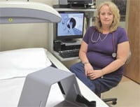
Lori Stoudenmire, a certified bone-density tech and medical assistant at Women’s Health Associates, is one of two techs at that facility who perform and read bone-densitometry scans.
“You can’t counsel women based just on their bone density,” she explained. “We have to ask questions that give us a true picture of what is really going on with their bones, not just how dense they are.”
Questions on the FRAX, she went on, include inquires about any past fractures of patients’ parents, which helps to identify genetic trends, and variables like race, age, height, weight, tobacco- and alcohol-consumption history, history of rheumatoid arthritis, nutritional and physical-injury history, steroid use (through medications, such as oral Prednisone), and medications (such as Prilosec for indigestion, which can stop absorption of calcium).
That information is calculated through the FRAX algorithm to come up with what are known as T-scores and Z-scores.
“T-scores compare the bone density of the patient to that of a young, healthy patient at the time of their peak bone mass, which is usually around age 23,” said Patel. “But another statistical measurement, called a Z-score, compares the bone density to one’s current age group.”
Patel told HCN that the T-scores are based on young, Caucasian male and female bone density, while the Z-scores are more population-specific, using different ethnicities, which, he admitted, makes it more difficult to qualify for risk-fracture analysis because not every ethnicity is accounted for and there are so many mixed races that it’s nearly impossible to segregate each ethnicity into a category.
Using the T-score (see chart) as the base for all analysis, the WHO has defined the following categories based on bone density in white women:
• Normal bone: T-score better than -1
• Osteopenia: T-score from -1 to -2.5
• Osteoporosis: T-score less than -2.5
Stoudenmire showed HCN the image of a healthy spine, which resembles a clean, straight chain of white rings. Conversely, a weaker, or osteoporotic, spine will have a mottled white and gray look and could have a slight or obvious curvature to it.
“We used to treat solely on bone density; if the patient had a T-score of minus 1.5 or worse, my inclination was to treat,” added Wool. “But we know that fracture risk is not based solely on bone density … there are lots of other factors.”
And those other factors are the gray area of bone disease where FRAX really comes into play.
Patel noted that, if a patient has osteoporosis, then the question of treatment isn’t an issue. “But if a patient has osteopenia, a lesser version of osteoporosis, and has a T-score of -1 to -2.5, the question is, ’should I treat the patient or should I wait?’ The FRAX really helps the clinician make that treatment decision.”
Quality Counts
While Wool focuses on preventative care and also advanced treatment of osteoporosis and fracture risk for those who really need it, another priority is to make sure insurance will cover those who need pharmacological therapy. And FRAX has helped him make that case for his patients.
“It’s helped me to convince insurance companies to pay for medication based on the FRAX analysis when a patient’s bone density wasn’t terrible, but their fracture risk was really high,” he explained. “It’s allowed me to override some of the insurance companies’ vetoes on some of the medications we use.”
On several occasions, Wool said, he’s seen insurance companies deny coverage for medication, and their reasoning is that the bone density isn’t bad enough to require the medication. Wool said he’ll respond with, “but look at her FRAX index; she’s at a high risk of a fracture — are you going to tell me she can’t have her medicine?” or words to that effect.
Wool added that the insurance companies are now accepting such arguments, but it will take more doctors like him to make sure they fight for their patients and get the correct information to each carrier.
Wool and Stoudenmire are also cognizant of what they call ’tech continuity.’
“When you go to the hospital to get the bone density, you get a different tech every time, a different radiologist every time, and interpretation of the report and quality may have a high variability,” Wool said. “Here, you get the same tech and the same person reading it, and it gets read in a way that I can interpret it. And I feel much more comfortable with my own reports than I do from an outside source, where I have to sort through the analysis.”
Wool was referring to the methodology between the GE enCORE and the Hologic software, which do not appear in the same layout for the final image and T-score analysis, but the goal is the same — to properly diagnose the potential for future bone fractures and osteoporosis through FRAX.
Both Wool and Stoudenmire said that the technician’s expertise can also make a big difference in a quality scan and the interpretation of the final FRAX information. The scan bed that Stoudenmire uses at Women’s Health Associates requires the patient to lie on her back with feet positioned in a foot frame, with no other positioning needed. Other densitometers require repositioning the patient, which can increase human error and decrease consistency in scans over the years.
“This is also where FRAX comes in to help offer more in-depth analysis to help interpret what is really going on,” said Patel.
Strong Future
As advances in technology continue, hospitals and medical offices are looking for more ways to correctly diagnose bone-fracture risk, which they know is on the rise due to the huge aging population.
Patel said that some Baystate locations are offering an additional analysis known as VFA, or vertebral fracture assessment, which also helps the clinician decide if a patient needs treatment or not. With a specific focus on the spine, said Patel, VFA provides a very low-dose X-ray of the thoracic and lumbar spine and can identify one small fracture in one level of the spine, which can highlight a sharply increased risk of future fracture in the patient. When used in conjunction with FRAX and bone densitometry, the most comprehensive diagnosis and path for treatment can be made.
Overall, this advancing technology is helping physicians and clinicians project years into the future and, ultimately, keep patients from reaching their breaking point — both literally and figuratively.


Comments are closed.